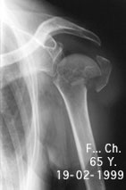
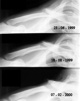
Shoulder fracture
Fracture in caput Valga on the humeral upper extremity.
Image n° 1: pre-operative (02 - 19 - 1999)
 |
|
Image n° 2:
post-operative
(02 - 21 - 1999)
|
|
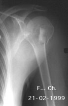 |
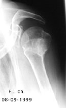
Image n° 3:
healing
(09 - 08 - 1999)
 |
|
Fracture of the collar bone
Atrophic non-union of the left collar bone: car accident
Image n° 1: pre-operative (08 - 31 - 2000)
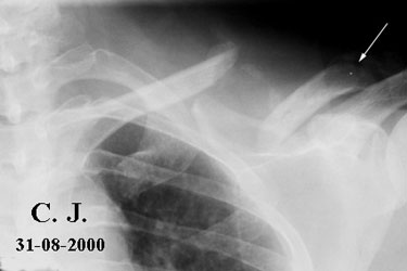
Note the large gap between the two fragments of the collar bone.
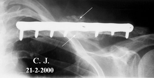
Image n° 2:
four months after surgery
(02 - 21 - 2000)
 |
|
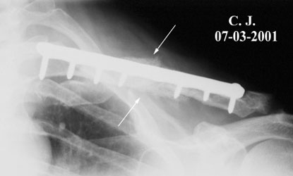
Image n° 3:
the fracture is healed
(03 - 07 - 2001)
|
|
 |
Atrophic non-union of the right collar bone
Clinical story:
![]() Y.F. is born in 1967. He is a professional teacher and tennis player. He is 15/20. He had a tongue's cancer which was treated locally. A bilateral removal of his neck's lymph node was performed, followed by radiotherapy (50 Gy on both cervical and collar fields).
Y.F. is born in 1967. He is a professional teacher and tennis player. He is 15/20. He had a tongue's cancer which was treated locally. A bilateral removal of his neck's lymph node was performed, followed by radiotherapy (50 Gy on both cervical and collar fields).
![]() He had a car accident in 1999 and breaks his right collar bone. He was treated orthopaedically. By times, no healing occurred.
He had a car accident in 1999 and breaks his right collar bone. He was treated orthopaedically. By times, no healing occurred.
![]() He could not play tennis anymore and his right arm became weaker. It lost two centimetres in diameter.
He could not play tennis anymore and his right arm became weaker. It lost two centimetres in diameter.
![]() No surgeons wanted to operate for two reasons they claim. The skin was atrophic due to the irradiation and they were afraid of a possible necrotic bone underneath. The man was hopeless.
No surgeons wanted to operate for two reasons they claim. The skin was atrophic due to the irradiation and they were afraid of a possible necrotic bone underneath. The man was hopeless.
![]() We had many operative possibilities to treat this case. We had a lot of discussion with the patient and his family and finally, we decided to remove the necrotic bone, to fix the fracture with a metallic plate and set a biomaterial graft.
We had many operative possibilities to treat this case. We had a lot of discussion with the patient and his family and finally, we decided to remove the necrotic bone, to fix the fracture with a metallic plate and set a biomaterial graft.
![]() We made an extemporaneous histological examination of the necrotic bone which reveals that « no signs of radiotherapic necrosis » was seen. It was confirmed three weeks later by a regular histological examination of bone samples.
We made an extemporaneous histological examination of the necrotic bone which reveals that « no signs of radiotherapic necrosis » was seen. It was confirmed three weeks later by a regular histological examination of bone samples.
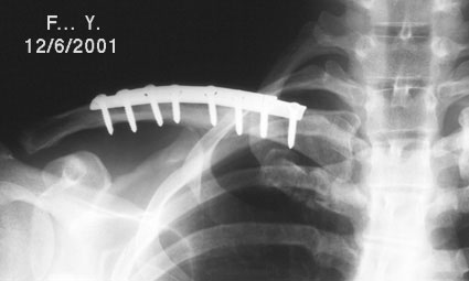
Image n° 1: Pre-operative X-rays
 |
|
Image n° 2: Bone healing
 |
|
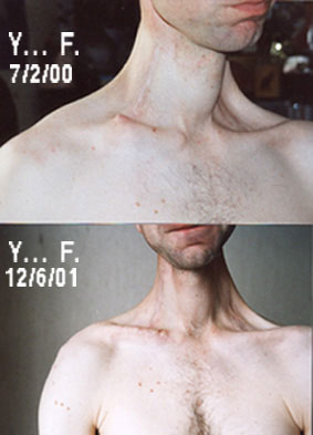
Image n° 3: Picture of both shoulders before and after surgery.
|
|
 |
Clinical follow-up:
![]() November 2005. No pain, no rehabilitation problems. YF returned to sport. He teaches and plays tennis. He is again 15.
November 2005. No pain, no rehabilitation problems. YF returned to sport. He teaches and plays tennis. He is again 15.
Conclusion:
![]() The clinical follow-up shows the good motor functions and recovery of all joints. For all of them, after more than eight years, there is no modification of the fracture site. The bone healing remains stable.
The clinical follow-up shows the good motor functions and recovery of all joints. For all of them, after more than eight years, there is no modification of the fracture site. The bone healing remains stable.
| Upper Limb pathology: Arm |