Colles' fracture
Radius fracture
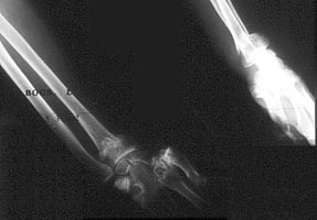
Image n° 1: pre-operative X-Rays

![]() The comminution (multiple fragments) of cortices (walls of bones) on this AP view is visible. The line between both extremities of radius and ulna is horizontal. There is a deformity of the wrist and hand which are deviated laterally. Note the subluxation of the joint between radius and ulna. There is a very little deformity on the lateral view. The decision to fix the fracture was done because of the severe arthrosis of the thumb's root. It was treated secondarily with prosthesis.
The comminution (multiple fragments) of cortices (walls of bones) on this AP view is visible. The line between both extremities of radius and ulna is horizontal. There is a deformity of the wrist and hand which are deviated laterally. Note the subluxation of the joint between radius and ulna. There is a very little deformity on the lateral view. The decision to fix the fracture was done because of the severe arthrosis of the thumb's root. It was treated secondarily with prosthesis.
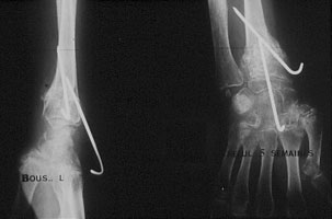
Image n° 2: X-Rays after 5 weeks

![]() Note the reconstruction of both cortices and the correction of the deformity. Natural coral granules were used in that case. We used as well two pins and an external split for 6 weeks.
Note the reconstruction of both cortices and the correction of the deformity. Natural coral granules were used in that case. We used as well two pins and an external split for 6 weeks.
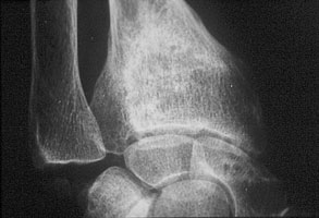
Image n° 3: Reconstruction of the lower part of the radius

![]() We can see the perfect remineralization of the wrist and the complete disappearing of the biomaterial. Note also the correction of the joint subluxation .
We can see the perfect remineralization of the wrist and the complete disappearing of the biomaterial. Note also the correction of the joint subluxation .