Measurement of bone loss
First: What is normal bone mass?
![]() At
the end of growth, towards the age of 20, skeletal growth is complete.
At
the end of growth, towards the age of 20, skeletal growth is complete.
A bone consists of an envelope made of compact bone, known as cortical
bone or cortex. Inside this envelope, at both ends of a long bone such
as the femur, is a bone made of cellular trabeculae: spongy bone. A graph
is produced showing the changes in Volume of these Bone Trabeculae –
Trabecular Bone Volume (TBV) – during human aging.
![]() At
the end of growth, TBV is at its highest level.
At
the end of growth, TBV is at its highest level.
As men age, it falls progressively. In women, it falls suddenly at menopause
and then continues to fall progressively as in men.
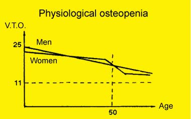
The two lines come together towards the age of 80.
This bone loss is natural. It is called physiological bone loss. Everyone loses bone as they age.
![]()
What is osteoporosis? What is osteoporotic disease?
![]() The
TBV provides a morphological analysis of bone and its physical structure.
The
TBV provides a morphological analysis of bone and its physical structure.
Chemically, bone consists of water, mineral salts, protein and fat.
![]() Bone Mineral Density
– BMD measures the quantity of mineral salts in bone. Unlike TBV
(trabecular bone volume) which measures only the trabeculae inside the
bone, osteodensitometry measures the mineral salts in both the envelope
and it's contents: in cortical and spongy bone.
Bone Mineral Density
– BMD measures the quantity of mineral salts in bone. Unlike TBV
(trabecular bone volume) which measures only the trabeculae inside the
bone, osteodensitometry measures the mineral salts in both the envelope
and it's contents: in cortical and spongy bone.
It can be evaluated in:
- the spine
- the hip
- the wrist and anckle.
![]()
What are the areas which are examined by bonedensitometry ?
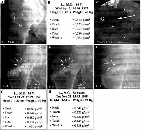
![]() Four
areas are examined in the upper femoral metaphysis:
Four
areas are examined in the upper femoral metaphysis:
- The greater trochanter
- The intertrochanteric zone
- The femoral neck
- The Ward's triangle
For further information on densitometers...
![]() This
exploration gives a quantitative measurement of mineral loss from bone
at four sites.
This
exploration gives a quantitative measurement of mineral loss from bone
at four sites.
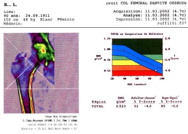
![]() Two
indexes have been defined to distinguish physiological osteopenia from
osteoporotic disease:
Two
indexes have been defined to distinguish physiological osteopenia from
osteoporotic disease:
- The T-score measures bone mineral quantity at its highest level. This
is the quantity found at a bone site in a young person.
- The Z-score measures bone mineral quantity of the subject examined compared
to a subject of the same age.
The osteoporotic disease is defined as a bone loss , 2,5 standard deviation
below the value observed in a normal young adult.
Global loss – whether from the envelope or the content – can
therefore reach 50, 60 and up to 90% of the maximum value.
In this particular case, there is only 0.035 g/cm² left.
The inside of the femoral neck is totally void of bone,
as can be seen in this osteoscopic image.
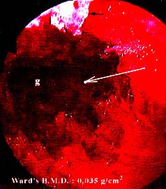
Histological section of an osteoporotic bone
![]() A
fracture threshold has been defined. It is the quantity of bone remaining,
below which there is a risk of fracture in the event of a fall.
A
fracture threshold has been defined. It is the quantity of bone remaining,
below which there is a risk of fracture in the event of a fall.
For the hip, this is 0.600 g/cm²..
The lower the threshold, the greater the theoretical risk of fracture.
see Articles: Cornell CN (1990) Management of fractures
in patients with osteoporosis
Orthop Clinical North Am 21 : 125-141
Barrios C and Coll (1993) Healing Complications after internal fixation
of trochanteric hip fractures :
The prognostic value of osteoporosis
J Orthop Trauma 7 : 438-442
![]() We
must emphasise a second point which interests trauma surgeons: the lower
the bone densitometry, the more complex the fracture is, the more difficult
its surgical stabilisation.
We
must emphasise a second point which interests trauma surgeons: the lower
the bone densitometry, the more complex the fracture is, the more difficult
its surgical stabilisation.
![]()