Arm fracture
Atrophic non-union of the humeral shaft
![]() Example of a humerus osteoarthritis.
Example of a humerus osteoarthritis.
Image n° 1: pre-operative (07 10 1991)
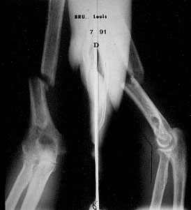
![]() Transverse fracture of the lower third. Note the lack of healing. No bone callus is seen. The medullary canal is closed.
Transverse fracture of the lower third. Note the lack of healing. No bone callus is seen. The medullary canal is closed.
Image n° 2: post-operative, day one (07 11 1991)

![]() Note the large amount of granules spread along the fracture site. A large decortication of the humeral shaft is done. The fracture is then firmly fixed by a metallic plate with screws. A simple splint is applied for six weeks before rehabilitation.
Note the large amount of granules spread along the fracture site. A large decortication of the humeral shaft is done. The fracture is then firmly fixed by a metallic plate with screws. A simple splint is applied for six weeks before rehabilitation.
Image n° 3: post-operative X-ray. Healing is complete.
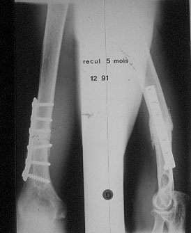
![]() The humerus heals in two months. On the radiography at 5 months, the reconstruction of both cortices is achieved. Note the homogeneous aspect of the bone. The granule's volume is decreasing and the bone callus is clearly seen.
The humerus heals in two months. On the radiography at 5 months, the reconstruction of both cortices is achieved. Note the homogeneous aspect of the bone. The granule's volume is decreasing and the bone callus is clearly seen.
| Upper Limb pathology: Forearm |