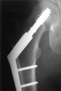
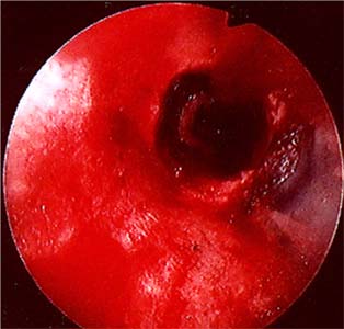
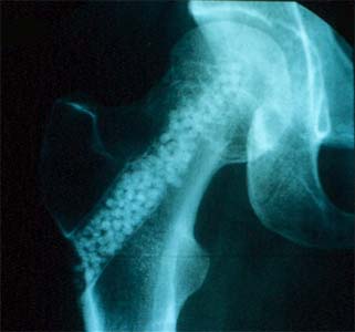
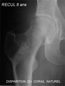
A 32 year old mother (born 22-11-1963) has severe osteoporosis. Will she have the courage to have another child? Mrs. C.S. is a 32 year old expectant mother. She is 8 and a half months pregnant when she slips in the bathroom, in July 1995. She breaks her right hip. She is taken to the Neuilly hospital, where she delivers a healthy baby by cesarean section. The same day, she is operated on her right hip. The fracture is just below the head of the femur and is secured with a screw plate. The bone densitometry of the left hip is very low. After doing a biological exam, the rheumatologist concludes that she has an algodystrophy of obstetrical origin. She is a woman of Israeli origin, who measures 1.70m and weighs 54kg. She lives in a well to do environment, has a supportive family and an active professional life. She is worried about the condition of her bones, and doesn't plan to have more children because of the fragility of her bones. In July 1997, two years later, she agrees to have the material removed. She also agrees to a graft of natural coral that was suggested to her for two reasons. The first is that in spite of the medical treatment she is receiving, the densitometry of the other hip remains low. The other is that, once the treatment has been fully explained to her and her family, she is convinced that the local insertion of calcium can improve the mechanical resistance of her bones by increasing her bone mass. The coral graft takes place in 1997, with a local osteoscopic evaluation during the operation . She agrees to have regular clinical, radiographic, densitometric and biological tests. The tests are therefore repeated regularly without fail from 1997 to 2005. Medically, Mrs. C.S. has never followed any particular treatment. She has a balanced diet. She has never taken anything but calcium and vitamin D supplements during the winter months. After two years, the osteosynthetic material is removed, and a coral graft is performed to fill the void created by the removal of the screw. A measure of bone densitometry of the broken hip is done in July 2005, when the coral is no longer visible. The conclusions of the exam are: the right cervical femoral BD is significantly higher than that of the side that hasn't been treated with coral.
THE FACTS YESTERDAY Mrs. C.S. at the age of 32 years, discovers, because of a fracture of her hip at the term of her first pregnancy, that she has fragile bones. She is worried about the future. Her family is very much aware of her problem and is also very concerned. Indeed, it is very unusual to take a bad fall in your bathroom and break your hip within days of delivering your first child... Mrs. C.S. is a tall young married woman, slim, who is very dynamic and strong-willed. She has a balanced diet, does sports on occasion (winter sports, swimming at the beach in the summer) and lives in pleasant environment. She also earns a good living. The fracture has profoundly disturbed her. The rheumatologist has concluded that she suffered from an algodystrophy of obstetrical origin. Since the delivery, the whole family lives in fear that another such incident will take place. She therefore changes her way of life. She stops doing any sports other than walking. She continues to work. The couple decides not to have any more children. THE FACTS TODAY Since 2004, Mrs C.S. leads a normal life. She has started doing sports again: she skis (on the most difficult slopes) plays tennis and walks a lot. She has had a second child. She is as active as before. She still does not take any medication. Would you dare? |
||||||||||||||