Fracture of the ankle
![]() The leg lower part is red. Blood and tar smear the skin except miraculously on the lateral side which is almost unhurt!
The leg lower part is red. Blood and tar smear the skin except miraculously on the lateral side which is almost unhurt!
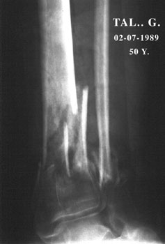 |
The first step is to clean the wound. The patient undergoing general anaesthesia, the surgeon patiently, meticulously, cleans the skin with iodine water. He removes inlaid stones, grass, earth and tar. Then he wraps the lower limb in sterile linen. During this time, he prepares the planning procedure. The first step is to restore the normal length. The clean lateral skin allows a plate metallic fixation of the fibula. Concerning the tibia, a big nail will be introduced through the medullary canal until it reaches the articular surface. It will give the proper and necessary alignment. |
Few months later
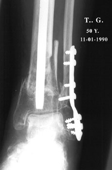 |
Positive points: The foot has been preserved. Negative points: There is a severe demineralization on both bones and on the foot. The lady does not complain and never asked for a proper realignment. |
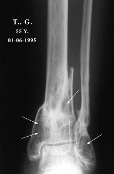 |
Five years later
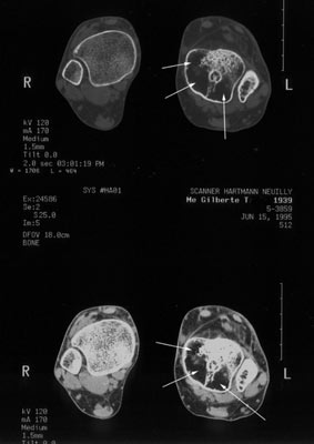
Pain happens during walking more and more frequently either during daytime or at night. Pain increases and is no longer relieved by pain-killer. The skin is warm and pink. No signs of infection. The joint is stiff and the foot mobile. The CT scan reveals a severe demineralization (arrows) either on the tibia and the fibula too. If it became necessary by time, in the future, to fix this joint, the technical problem will be difficult because of that bone loss. A graft will be necessary for sure. |
Another view of the tibia (CT scan)
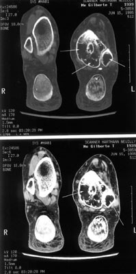 |
Why should we wait? There are two reasons not to waste time. I°- The functional joint disability. Another question is: why the joint is so painful? The fracture is healed, the metallic implants were removed. The vicious callus of the tibial lower extremity? The joint stiffness? The severe bone mass loss (compared both right and left side on the CT scan)? Whatever the cause, a bone graft would be necessary if any surgical procedure is required. |
Few weeks after the bone graft
 |
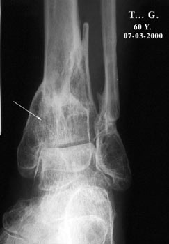
Natural coral did you say! Yes, but why? According to our knowledge, natural coral is a bone substitute. It becomes by time a new bone, part of our skeleton. Here, it is a thick and strong cortical bone; there it is similar to the bee cells. By time, as a matter of fact, it undergoes through mechanical transformations like a normal bone, whatever the strains. Here, if it is placed around a cortical wall, the histological samples will show a Haversian structure. There if it fills a cancellous defect, it becomes a cancellous bone. Natural coral did you say? OK but how? AP view : |
 |
What about the lateral view? What do you think? It is not so bad, isn't it? |
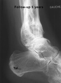 |
Five years later Natural coral disappeared. It is now the lady's bone. Did you say…Magic? |
| Previous page |
| Lower Limb pathology: Calcaneeum |