Tibia fracture
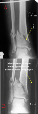
Atrophic non-union of the tibia 4 months after a road car accident
 |
This 82 years old lady was hit by a car. She lost consciousness for three weeks. Her skin's leg was open at the level's fracture and needed a large muscular and cutaneous flap to cover the exposit bone. An external metallic fixation was put through the skin either in the upper part of the non broken tibia and below in the calcaneum (heel bone). No bone callus formation is seen (picture A). The fracture site changed as soon as we tried to loosen some screws (photo B). The procedure was to put in close contact both parts of the tibia (i), to bring locally a biological material (natural coral), and to fix strongly the fracture site. |
Remarks: What is an ankle joint?
Let us give some technical explanations.
![]() As we all know that there are an upper part and a lower part in a room, there are two heels.
As we all know that there are an upper part and a lower part in a room, there are two heels.
![]() The upper heel close to the leg and the lower heel close to the foot.
The upper heel close to the leg and the lower heel close to the foot.
![]() I° - the bone which is six to ten centimetres above the joint needs always a long time to heel. Three months at least, four months or even more!
I° - the bone which is six to ten centimetres above the joint needs always a long time to heel. Three months at least, four months or even more!
![]() II° - The bone between one to four centimetres close to the joint heals more easily… unless certain circumstances do not allow the bone consolidation. One of them is a bone mass loss such as severe osteoporosis for example.
II° - The bone between one to four centimetres close to the joint heals more easily… unless certain circumstances do not allow the bone consolidation. One of them is a bone mass loss such as severe osteoporosis for example.
The middle part of the tibia sinks like a pile in the osteoporotic lower extremity.
 |
A long bone is a hollow tube. The great tibial axis is almost properly lined-up (A) on this lateral view. But, as soon as the metallic fixation loosens (B). I° -the fragment A penetrates the fragment B. II° - Some small cortical fragments penetrates the hollow tube. III° - The cancellous bone of the fragment B was highly compressed during the bump. |
Is it a good job?
 |
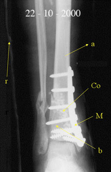
Fragments A and B are in close contact. The cancellous bone gap is filled with the biomaterial (Co). The tibia is anatomically rebuilt and the long axis is perpendicular to the ground. The metallic plate (M) fixation is mechanically strong. A resin is applied for six weeks. Total weight-bearing is not allowed during six weeks. |
What do you say about consolidation?
 |
The resin is removed after six weeks. The rehabilitation of the joint begins... Three months after, X-Ray shows a bone healing sufficient to allow total weight-bearing with two crutches. One month after, walking without crutches is allowed. |
 |
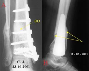
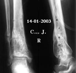
One month later, on the X-rays: Beads of natural coral disappear (A). Reconstruction of walls is done (B). Both fracture sites are healed. The ankle joint is mobile. The bone non-homogeneous aspect will disappear by time. |
| Next page |