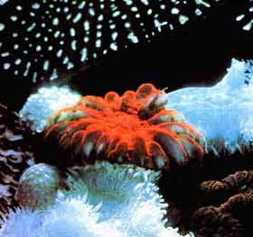
Coral
What is biological material? Is natural coral a biomaterial?
![]() Very
briefly, a biological material is a product which, inserted into the body,
is not rejected. It does not generate immune reactions. It is "accepted"
by the body.
Very
briefly, a biological material is a product which, inserted into the body,
is not rejected. It does not generate immune reactions. It is "accepted"
by the body.
What are the characteristics of natural coral?
General:
![]() Jean André
PEYSONNEL, an 18th century French surgeon, determined the fact that corals
were animals: coral polyps. In their tissues they maintain single-celled
algae, zooxanthellae, which use photosynthesis to produce the elements
which are essential to the corals. They also remove toxic residues. Chlorophyll,
combined with various pigments thus helps give corals their different
colours.
Jean André
PEYSONNEL, an 18th century French surgeon, determined the fact that corals
were animals: coral polyps. In their tissues they maintain single-celled
algae, zooxanthellae, which use photosynthesis to produce the elements
which are essential to the corals. They also remove toxic residues. Chlorophyll,
combined with various pigments thus helps give corals their different
colours.
For
further information: a Natural coral production unit
![]()
Physical characteristics of coral:
![]() This material, of
natural origin, is a Calcium carbonate compound in its crystalline phase:
aragonite. It displays great architectural regularity, with open pores
allowing fluids to circulate freely inside the skeleton.
This material, of
natural origin, is a Calcium carbonate compound in its crystalline phase:
aragonite. It displays great architectural regularity, with open pores
allowing fluids to circulate freely inside the skeleton.
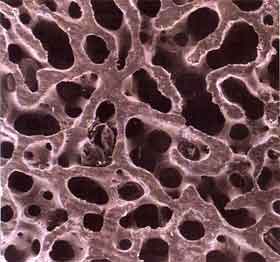 Cross-section of human spongy bone |
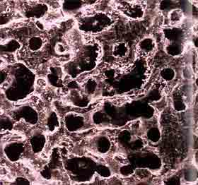 Cross-section of coral porites displaying 50% porosity |
| Here you can see the similarity between the two structures | |
![]() This
porosity varies according to the species of coral under consideration.
The volume of porosity, pore interconnection, regularity and diameter
(150 µm on average) all mean that, once implanted in the bone tissue,
there is total and rapid invasion of the graft by blood or bone marrow
cells, vascularisation is established (insert here calculations of pore
areas and volumes). The coral architecture provides an exceptional surface
for exchanges between the biomaterial and the bone..
This
porosity varies according to the species of coral under consideration.
The volume of porosity, pore interconnection, regularity and diameter
(150 µm on average) all mean that, once implanted in the bone tissue,
there is total and rapid invasion of the graft by blood or bone marrow
cells, vascularisation is established (insert here calculations of pore
areas and volumes). The coral architecture provides an exceptional surface
for exchanges between the biomaterial and the bone..
![]()
Biomechanical characteristics of
coral:
![]() The mechanical characteristics
of coral depend on the haemodynamic constraints it is under and the organisation
and volume of its porosity. They therefore differ according to the type
and species under consideration.
The mechanical characteristics
of coral depend on the haemodynamic constraints it is under and the organisation
and volume of its porosity. They therefore differ according to the type
and species under consideration.
For further information on the mechanical characteristics of coral
![]()
Chemical characteristics of coral
:
![]() In the book: Les
coraux, B. Robin, C. Petron and C. Rives and coll. have shown that the
primary phase in the development of the coral skeleton resembles mammalian
osteogenesis in its fundamental mechanisms. Corals are essentially composed
of mineral elements but also several appropriate amino acids which
should be eliminated, or at least reduced as much as possible - using
specific purification processes such as supercritical fluids to
avoid, minimise and/or remove any harmful immune reactions.
In the book: Les
coraux, B. Robin, C. Petron and C. Rives and coll. have shown that the
primary phase in the development of the coral skeleton resembles mammalian
osteogenesis in its fundamental mechanisms. Corals are essentially composed
of mineral elements but also several appropriate amino acids which
should be eliminated, or at least reduced as much as possible - using
specific purification processes such as supercritical fluids to
avoid, minimise and/or remove any harmful immune reactions.
![]() There
are differences between coral and fresh bone. The mineral phase in particular
2/3 of bone composition which is essentially in the form
of calcium phosphate. Although the organic phase is important in bone
1/3 of its composition it is reduced to a very small proportion
of amino acids in coral. However these should be eliminated to avoid any
risk of immune reaction.
There
are differences between coral and fresh bone. The mineral phase in particular
2/3 of bone composition which is essentially in the form
of calcium phosphate. Although the organic phase is important in bone
1/3 of its composition it is reduced to a very small proportion
of amino acids in coral. However these should be eliminated to avoid any
risk of immune reaction.
![]() There
are analogies: Two have specific action. Strontium is involved in the
growth of bone crystal. Fluorine increases bone formation in spongy bone,
in small doses and has the opposite effect on the bone walls at high concentration.
It acts by stimulating osteoblast precursor cells.
There
are analogies: Two have specific action. Strontium is involved in the
growth of bone crystal. Fluorine increases bone formation in spongy bone,
in small doses and has the opposite effect on the bone walls at high concentration.
It acts by stimulating osteoblast precursor cells.
For further information on the chemical characteristics of coral
![]()
Biocompatibility :
![]() Tests
were performed on rats, rabbits, sheep, pigs and dogs. They concerned
the behaviour of tissues coming into contact with the material in various
implantation sites: subcutaneous, intramuscular, intraosseous, subperiosteal
and alveolodental.
Tests
were performed on rats, rabbits, sheep, pigs and dogs. They concerned
the behaviour of tissues coming into contact with the material in various
implantation sites: subcutaneous, intramuscular, intraosseous, subperiosteal
and alveolodental.
![]() No
acute or chronic inflammatory reactions were observed, nor any granulocytic
infectious reaction, nor any rejection with proliferation of round cells
or fibrous encapsulation. No tissue concerned showed any immune reaction
against the biomaterial. Very good tolerance was observed in all cases.
No
acute or chronic inflammatory reactions were observed, nor any granulocytic
infectious reaction, nor any rejection with proliferation of round cells
or fibrous encapsulation. No tissue concerned showed any immune reaction
against the biomaterial. Very good tolerance was observed in all cases.
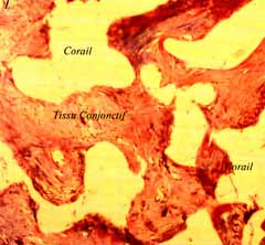 Coral in skin |
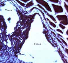 Coral in muscle |
 Coral in bone |
 Coral in bone |
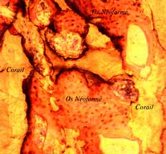
 Coral in neoformed bone |
 Coral in neoformed bone |
![]()
Bioresorbability and coral in animals :
![]() Filling
a collapsed spongy bone in dogs with a fragment of coral implanted
in one end of the bone shows almost total resorption of the biomaterial
in six weeks and its replacement by bone tissue.
Filling
a collapsed spongy bone in dogs with a fragment of coral implanted
in one end of the bone shows almost total resorption of the biomaterial
in six weeks and its replacement by bone tissue.
![]() Histological
studies have shown, particularly in dogs and pigs, the presence of numerous
osteoclasts in contact with the coral during resorption. Carbonic anhydrase
was revealed in the osteoclasts. The involvement of carbonic anhydrase
in the destruction of carbonate substrates has been studied since 1969
and the demonstration of this role proved by the slowing of destruction
after administration of acetazolamide, a specific carbonic anhydrase inhibitor
.
Histological
studies have shown, particularly in dogs and pigs, the presence of numerous
osteoclasts in contact with the coral during resorption. Carbonic anhydrase
was revealed in the osteoclasts. The involvement of carbonic anhydrase
in the destruction of carbonate substrates has been studied since 1969
and the demonstration of this role proved by the slowing of destruction
after administration of acetazolamide, a specific carbonic anhydrase inhibitor
.
![]() To
verify the hypothesis of this enzyme's role in the destruction of coral
carbonate skeletons, 10 femoral transcortical resections involving replacement
by this biomaterial have been performed on animals treated with acetazolamide.
To
verify the hypothesis of this enzyme's role in the destruction of coral
carbonate skeletons, 10 femoral transcortical resections involving replacement
by this biomaterial have been performed on animals treated with acetazolamide.
In comparison with untreated subjects, a definite slowing in the resorption
of implanted coral was noted. This was accompanied by bone necrosis on
the edge of the graft, which tended to spread to the entire bone. These
resections were not consolidated, even one year later and all led to pseudarthrosis.
The resorption of coral carbonate skeleton is, at least partly, due to
the carbonic anhydrase in osteoclasts.
![]()
Histology:
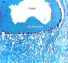
![]() The transformation
of coral into bone takes place in several successive phases. These are
embedded as the calcification front advances.
The transformation
of coral into bone takes place in several successive phases. These are
embedded as the calcification front advances.
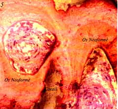
| Experiments led, histologically, to the revelation of five phases, recorded consistently, which are successive and embedded as the resorption front and calcification fronts advance. | |
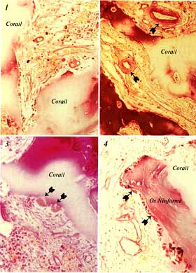 Iconography Group "Formation et Destruction des Tissus calcifiés" (Formation and destruction of calcified tissues) University Paris VII Biology and Genetics research unit Paris FRANCE |
Phase 1 (Image 1) : x 200 PAS colouring. Preparation as demineralised tissue. The coral is partly demineralised and appears to be translucid. The entire porosity is invaded by the coral implanted in the femoral diaphysis of a dog. Phase 2 (Image 2) : x 160 Masson trichrome stain. Preparation as demineralised tissue, but the coral is only partly demineralised. In this image, two arterioles can clearly be seen (arrows) and the shells of their walls can be clearly differentiated. Phase 3 (Image 3) : x 1200 Haematoxylin-eosin stain. Preparation as demineralised tissue. The coral is only partly decalcified and appears as a whitish area. Two osteoclasts are on the edge of the coral, their brush-like edges directed towards the material to be resorbed (arrows) . Phase 4 (Image 4) : x 180 Haematoxylin-eosin stain. Preparation as decalcified tissue, with the coral only partly decalcified. The neoformed bone is displayed as deep pink, with the first osteoblastic layer laid directly on the coral: successive layers will gradually thicken the new trabeculae (arrows). |
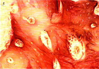 |
Phase 5 (Image 5) : x 200 Masson's trichrome stain. Preparation as demineralised tissue. Cortical bone site of implantation of a fragment of CORAL in a dog's femur, after 18 months. At the end of the process, BIOCORAL© has been totally resorbed, the neoformed bone tissue which initially formed the coral architecture has been remodelled as haversian tissue. Iconography: |
![]()
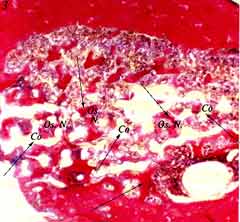
The resorption process has been studied in dogs and pigs.
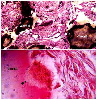 |
Image 1 x 1000 - Paragon stain Implantation in a pig's metaphysis, 4 weeks after surgery. The coral is dark brown, the edges fringed with osteoclastic action (arrows). The non-demineralised preparation technique does not allow accurate identification of cells. This is done on decalcified preparation. Iconography: L.R.O. U.A. CNRS 1161 Paris FRANCE Image 2 x 1000 - Haematoxylin-eosin stain The osteoclast, with its brush-like border, is clearly visible, directly against the coral and hollowing a resorption lacuna (arrow). Iconography: |
![]()
| Next page |