Reconstruction of spongy bone
using coral
In humans: A biological material must have the following characteristics:
It must be bioresorbable (gradual complete disappearance)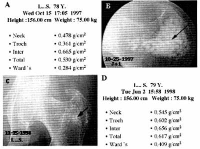
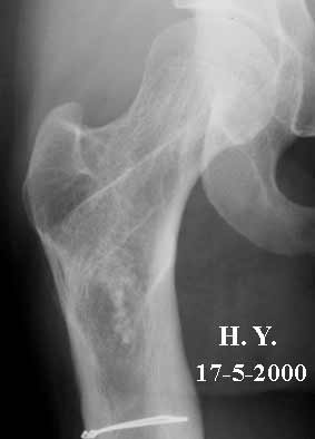
It can be seen in this image that the coral implanted in the greater trochanter
has disappeared just over a year later.
 |
 |
 |
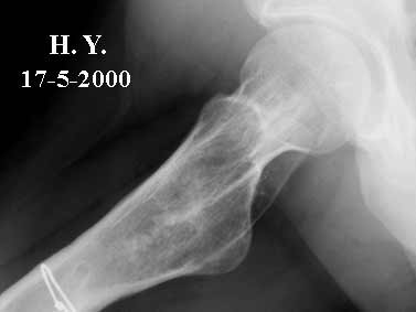 |
| Note the complete disappearance of the natural coral in two years | |
![]()
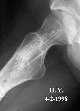
You can see that it is impossible to distinguish the implanted zone from
the non-implanted bone
in the spongy bone of this patient's femur after disappearance of the
biomaterial.
![]()
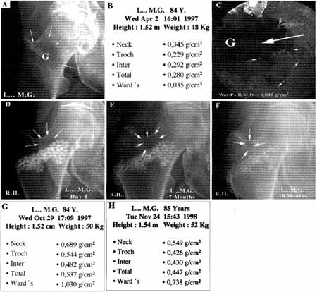
In this case, there is a steep increase in BMD.
![]()
![]() Verification
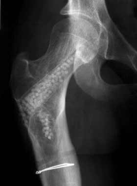
on an anatomical specimen must show the presence of biomaterial
if it is only partly resorbed. This is shown here on a CT scan of an anatomical
specimen. Note the layout in the form of trabeculae forming an arch, which
is characteristic of normal bone, shown here in an implanted hip.
Verification
on an anatomical specimen must show the presence of biomaterial
if it is only partly resorbed. This is shown here on a CT scan of an anatomical
specimen. Note the layout in the form of trabeculae forming an arch, which
is characteristic of normal bone, shown here in an implanted hip.
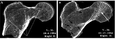 Right hip not implanted: note the size of the bony void, the complete disappearance of bone trabeculae. The remaining bone trabeculae are the site of microfractures (arrows on left slide) which have been described by Maurice Michel Forest. |
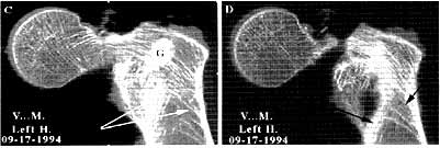 Implanted left hip : note the partial persistence of the coral (on left slide). The bone reconstruction with redevelopment of good arched trabecular characteristic of the anatomy of the upper end of the femur. |
![]()
![]() The
biomaterial must behave like a normal bone, i.e. reduce
regularly with time in the same proportions as the physiological reduction.
BMD measurements in the following table confirm this reduction with longitudinal
monitoring for more than 6 years. The BMD and date in months (m) or years
(y) is given for each post-operative examination.
The
biomaterial must behave like a normal bone, i.e. reduce
regularly with time in the same proportions as the physiological reduction.
BMD measurements in the following table confirm this reduction with longitudinal
monitoring for more than 6 years. The BMD and date in months (m) or years
(y) is given for each post-operative examination.
| Patient | Age | Date of graft | Post-Op 1 | Post-Op 2 | Post-Op 3 | Post-Op 4 | Post-Op 5 | Post-Op 6 | ||||||
| C.J. | 66 | 3/07/92 | 0,559 | 10 m | 0,541 | 2 y | - | - | - | - | - | - | - | - |
| T.S. | 76 | 28/01/92 | 0,648 | 2 m | 0,641 | 3 m | - | - | - | - | - | - | - | - |
| M.M. | 79 | 2/10/92 | 0,566 | 4 m | 0,852 | 7 m | 0,549 | 20 m | 0,416 | 40 m | - | - | - | - |
| M.O. | 80 | 24/07/92 | 0,853 | 6 m | 0,852 | 1 y | 0,742 | 2 y | 0,667 | 4 y | 0,645 | 5 y | 0,636 | 6 y |
| V.L. | 83 | 2/10/92 | 0,408 | 3 m | 0,494 | 1 y | 0,498 | 2 y | - | - | - | - | - | - |
| O.G. | 84 | 4/12/92 | 0,597 | 3 m | 0,589 | 6 m | 0,596 | 1 y | 0,588 | 18 m | - | - | - | - |
| V.B. | 87 | 27/11/92 | 0,868 | 3 m | 0,803 | 2 y | - | - | - | - | - | - | - | - |
| G.A. | 94 | 27/10/92 | 0,806 | 3 m | 0,773 | 6 m | 0,691 | 1 y | 0,668 | 14 m | 0,622 | 22 m | 0,541 | 54 m |
![]()
Histology of natural coral in a bone medium
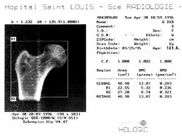
![]() This
patient died at the age of 101 and had suffered a fracture of the femoral
neck at the age of 95.
This
patient died at the age of 101 and had suffered a fracture of the femoral
neck at the age of 95.
![]() She
no longer left her room and was bedridden because of an inoperable brain
tumour. Examination of both hips 6 years after death revealed the transformation
of the coral in the fractured hip which had received a graft.
She
no longer left her room and was bedridden because of an inoperable brain
tumour. Examination of both hips 6 years after death revealed the transformation
of the coral in the fractured hip which had received a graft.
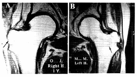
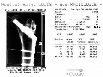
Bone densitometry performed on post-mortem samples from both femurs showed
the increase in bone mineral density on the implanted side. The bone mineral
density of the left hip reflects the bone's pre-existing bone mineral
density plus that of the bone neoformed from coral and that of the remaining
coral, whereas the bone mineral density of the right hip reflects the
remaining bone.
![]()
![]()
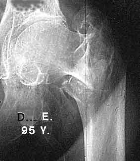
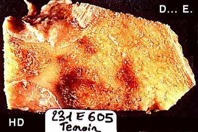
![]() On
the right hand side, the section of femur shows almost complete
disappearance of the bone lamellae, replaced by fat. There is no architectural
organisation remaining.
On
the right hand side, the section of femur shows almost complete
disappearance of the bone lamellae, replaced by fat. There is no architectural
organisation remaining.
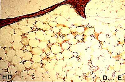
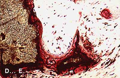
![]() Microscopic
examination revealed the same phenomenon, i.e. the extreme rarity of bone
lamellae here a bone lamella is coloured red-brown. It has lost
all connection with the adjacent bone lamellae. It is surrounded by adipose
cells. The more advanced the osteoporotic disease, the fewer bone lamellae
there are.
Microscopic
examination revealed the same phenomenon, i.e. the extreme rarity of bone
lamellae here a bone lamella is coloured red-brown. It has lost
all connection with the adjacent bone lamellae. It is surrounded by adipose
cells. The more advanced the osteoporotic disease, the fewer bone lamellae
there are.
![]()
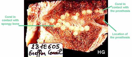
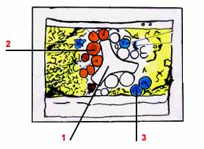
![]() On
the left-hand side, examination of bone cross-section with the
naked eye shows, firstly, an empty space in the middle corresponding to
surgical material surrounded by several spheres, some of which are clearly
visible and in contact with the material, with others more or less destroyed,
in contact with the adjacent bone.
On
the left-hand side, examination of bone cross-section with the
naked eye shows, firstly, an empty space in the middle corresponding to
surgical material surrounded by several spheres, some of which are clearly
visible and in contact with the material, with others more or less destroyed,
in contact with the adjacent bone.
 Cross-section parallel with the main axis of the femoral neck |
|
 Section perpendicular to the axis of the femoral neck |
![]()
![]() The
natural coral spheres have remained intact because, in contact with the
metal which has no vascularisation, they have been surrounded by tissue
with no vessels a sort of tissue packing.
The
natural coral spheres have remained intact because, in contact with the
metal which has no vascularisation, they have been surrounded by tissue
with no vessels a sort of tissue packing.
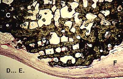
![]() On
the other hand, the coral in contact with the bone and vessels attached
to it, has been colonised by bone cells which have resorbed the biomaterial.
On
the other hand, the coral in contact with the bone and vessels attached
to it, has been colonised by bone cells which have resorbed the biomaterial.
Work performed by the Biomatech Laboratory
![]()
| Last page | Next page |