Reconstruction of cortical bone
using coral
![]() We
have just seen the reconstruction of spongy bone at the ends of a long
bone, using natural coral. Now let's see how to rebuild the wall (cortical
bone) of a long bone.
We
have just seen the reconstruction of spongy bone at the ends of a long
bone, using natural coral. Now let's see how to rebuild the wall (cortical
bone) of a long bone.
![]() Here's
an example of a young man, 20 years old, who was injured in a road accident
(long-term follow-up for 10 years).
Here's
an example of a young man, 20 years old, who was injured in a road accident
(long-term follow-up for 10 years).
Patient's clinical history:
![]() After
many problems, he saw the doctor about the delay in consolidation of his
femur. He had two metal devices (external fixators), one in front and
one on the side of the thigh. The knee was locked and stiff in extension.
The thigh bone (femur) was not strong. There were several centimetres
missing between the two ends. The ends were stripped and white. In short,
this bone had to be consolidated by the insertion of material. The classic
technique consists of taking a fragment of the patient's bone from the
pelvis (a graft) and inserting it so that it fills the void and consolidates
the bone.
After
many problems, he saw the doctor about the delay in consolidation of his
femur. He had two metal devices (external fixators), one in front and
one on the side of the thigh. The knee was locked and stiff in extension.
The thigh bone (femur) was not strong. There were several centimetres
missing between the two ends. The ends were stripped and white. In short,
this bone had to be consolidated by the insertion of material. The classic
technique consists of taking a fragment of the patient's bone from the
pelvis (a graft) and inserting it so that it fills the void and consolidates
the bone.
![]() This excellent technique is tried and tested and practised by most traumatologists.
If we can accept the idea that a biomaterial behaves like an immunologically
compatible bone graft, it should be possible to obtain the same result
but with a distinct advantage. We are not obliged to take a bone graft.
We don't have to perform another operation which is always painful.
This excellent technique is tried and tested and practised by most traumatologists.
If we can accept the idea that a biomaterial behaves like an immunologically
compatible bone graft, it should be possible to obtain the same result
but with a distinct advantage. We are not obliged to take a bone graft.
We don't have to perform another operation which is always painful.
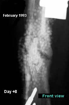
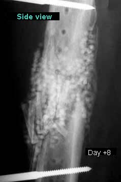
![]() The
coral implantation was performed. The coral spheres on all four sides
of the bone wall were counted.
The
coral implantation was performed. The coral spheres on all four sides
of the bone wall were counted.
Counting spheres
|
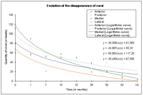
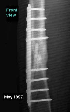
![]() A
parallel was established between the disappearance of the spheres and
the formation of the bony callus.
A
parallel was established between the disappearance of the spheres and
the formation of the bony callus.
Compared evolution of callus
development and disappearance of the coral
|
||||||||||||||||||||||||||||||||||||||||||||||||||||||||||||||||||||||||||||||||||||||||||||||||||||||||||||||||||||||||||||||||||||||||||||||||||||||||||||||||||||||||||||||||||||||||||||||||||||||||||||||||||||||||||||||||||||||||||||||||||||||||
![]()
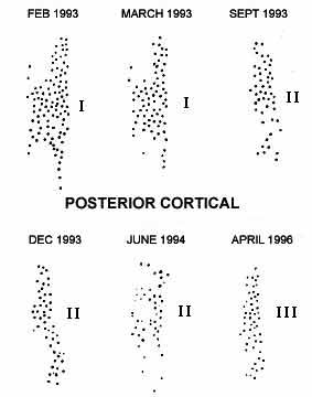
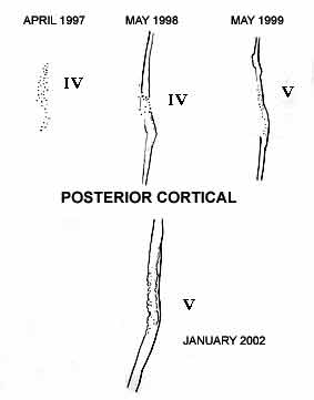
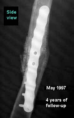
![]() A
complete and gradual reconstruction of the femoral walls (container) took
place. Reconstruction involved five phases (numbered in Roman numerals).
A
complete and gradual reconstruction of the femoral walls (container) took
place. Reconstruction involved five phases (numbered in Roman numerals).
I: FIRST BIORESORBABILITY II: ALIGNMENT IN A COLUMNAR RIDGE II: REDUCING THE DIAMETER OF THE COLUMNS III: REMODELLING THE COLUMNAR RIDGE IV: SECOND RESORBABILITY V: CORTICALISATION AND CORTICAL THICKENING |
![]()
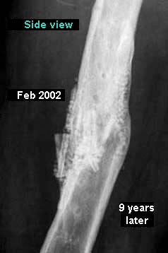
![]() X-rays
copies on tracing paper shows the progress of reconstruction of the wall
the container.
X-rays
copies on tracing paper shows the progress of reconstruction of the wall
the container.
 |
 |
![]() We
trace the logarithmic curves of the coral disappearance.
We
trace the logarithmic curves of the coral disappearance.
![]()
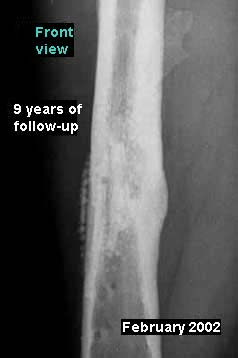
![]() A
magnificent femoral diaphysis (part of the femur between hip and knee)
is reconstructed before your eyes.
A
magnificent femoral diaphysis (part of the femur between hip and knee)
is reconstructed before your eyes.
  |
  |
  |
![]()
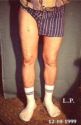
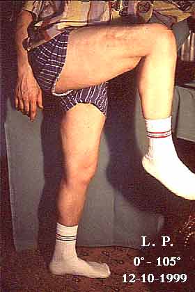
![]() The
result relative to knee stability and mobility is clear (mobility: 0-105°).
The
result relative to knee stability and mobility is clear (mobility: 0-105°).
 |
 |
![]()
| Previous page |