Preventive surgical treatment
What are the first results?
![]() Since 1990, 45 people
suffering from femoral neck fractures have agreed to have the opposite
hip grafted preventively. Some of these died without fractures. Two suffered
fractures within two months of grafting for reasons described above.
Since 1990, 45 people
suffering from femoral neck fractures have agreed to have the opposite
hip grafted preventively. Some of these died without fractures. Two suffered
fractures within two months of grafting for reasons described above.
The others are monitored regularly, either by telephone or in the surgery.
An article is being prepared to discuss the grounds for this treatment.
So far there have been no complications rejection, pain, weakening,
infection, limping etc. There have not been any fractures in these patients.
On the other hand, two patients who, for various reasons, had refused
the graft, suffered fractures within six months of the first fracture.
Here are a few examples:
1 - Mrs A.C.
![]() Born
in 1926. She is a tall woman (1m 78) elegant and very positive in outlook.
She works as a company chairman and knows what she is worth. At the age
of 38 she had part of her uterus and her two ovaries removed. To prevent
osteoporosis developing secondarily to the loss of hormone secretions
and because of her gynaecological problems, she could not take hormone
replacement therapy. She was therefore treated from 1992 to 1996 with
biphosphonates, calcium and vitamin D. A bone densitometry examination
made by the same operator with the same machine, showed BMD stabilisation
of the greater trochanter from 1992 to 1994. Treatment was therefore continued.
A further densitometry performed in 1996, on the other hand, shows major
loss of BMD. She had lost nearly a quarter of her bone mineralogy in two
and a half years.
Born
in 1926. She is a tall woman (1m 78) elegant and very positive in outlook.
She works as a company chairman and knows what she is worth. At the age
of 38 she had part of her uterus and her two ovaries removed. To prevent
osteoporosis developing secondarily to the loss of hormone secretions
and because of her gynaecological problems, she could not take hormone
replacement therapy. She was therefore treated from 1992 to 1996 with
biphosphonates, calcium and vitamin D. A bone densitometry examination
made by the same operator with the same machine, showed BMD stabilisation
of the greater trochanter from 1992 to 1994. Treatment was therefore continued.
A further densitometry performed in 1996, on the other hand, shows major
loss of BMD. She had lost nearly a quarter of her bone mineralogy in two
and a half years.
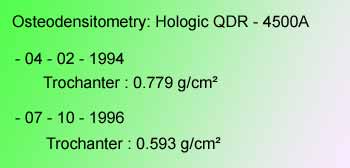
Preoperative BMD
Note the loss of 225 mg/cm² in four years
![]() During
the examination of October 1996, it emerged that she was suffering pain
in her left hip following mechanical effort. She had lost 3 cm in height.
There was marked demineralisation on the X-rays. A natural coral filling-graft
was performed at the end of March 1997.
During
the examination of October 1996, it emerged that she was suffering pain
in her left hip following mechanical effort. She had lost 3 cm in height.
There was marked demineralisation on the X-rays. A natural coral filling-graft
was performed at the end of March 1997.
Five years later, in June 2002, an X-ray confirmed the almost total disappearance
of the graft.
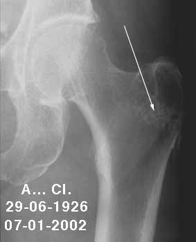
Filling-graft: disappearance of the coral
![]() The
bone densitometry performed at the end of December 2001 shows conservation
of the BMD at a level almost identical to that of three years previously.
The
bone densitometry performed at the end of December 2001 shows conservation
of the BMD at a level almost identical to that of three years previously.
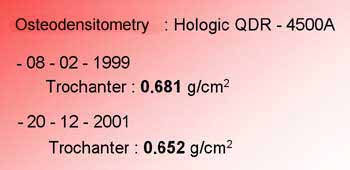
Note the stability of BMD five years after the graft
![]() In
this case, preventive medical treatment did not prevent a 0.126 g/cm²
loss in BMD in two years. Surgical treatment showed that, five years after
the graft, BMD was stable with a gain of 0.59 g/cm².
In
this case, preventive medical treatment did not prevent a 0.126 g/cm²
loss in BMD in two years. Surgical treatment showed that, five years after
the graft, BMD was stable with a gain of 0.59 g/cm².
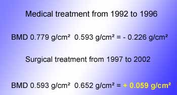
Comparison of BMD after medical and surgical treatment
![]()
2 - Mrs B.M.
![]() Falling
a fall from her own height, this lady suffered a fracture on 18th
August 1996, on the left side. She had surgery the next day, with a one-piece
nail-plate combined with a natural coral filling graft. Full weight-bearing
with two crutches was achieved ten days later. Full weight-bearing with
a single stick was achieved on the 45th day, the fracture being
consolidated with respect to the bone. This hip was weakened by hemiplegic
sequelae.
Falling
a fall from her own height, this lady suffered a fracture on 18th
August 1996, on the left side. She had surgery the next day, with a one-piece
nail-plate combined with a natural coral filling graft. Full weight-bearing
with two crutches was achieved ten days later. Full weight-bearing with
a single stick was achieved on the 45th day, the fracture being
consolidated with respect to the bone. This hip was weakened by hemiplegic
sequelae.
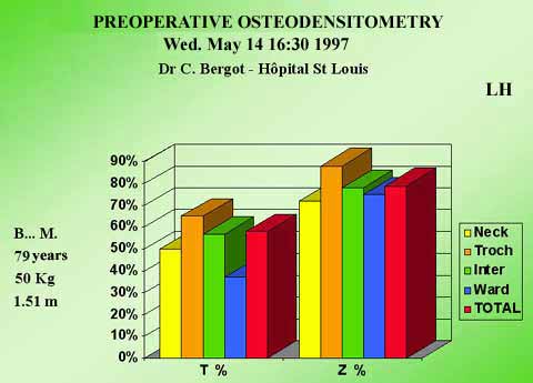
![]() An
osteodensitometry was performed nine months later in May 1997.
An
osteodensitometry was performed nine months later in May 1997.
![]()
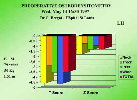
![]() It
shows a global loss of nearly half the BMD. The greatest loss is in the
central part of the bone (Ward's triangle).
It
shows a global loss of nearly half the BMD. The greatest loss is in the
central part of the bone (Ward's triangle).
The coral graft was performed during the next ten days. It filled most
of the Ward's triangle..
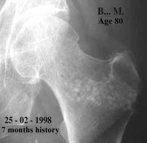
![]() An
X-ray during the seventh month shows the disappearance of nearly 80% of
the spheres.
An
X-ray during the seventh month shows the disappearance of nearly 80% of
the spheres.
 |
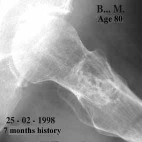 |
| Left hip front view | Left hip side view |
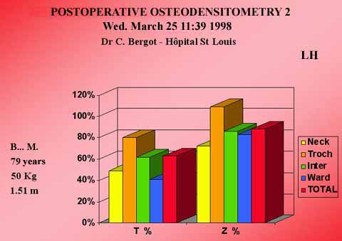
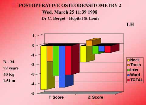
![]() The
second densitometry performed in March 1998 one year later
shows that there is no change in BMD either in the femoral neck or Ward's
triangle. On the other hand, there is a significant increase in the greater
trochanter (0.110 g/cm²), a zone which had not been grafted.
The
second densitometry performed in March 1998 one year later
shows that there is no change in BMD either in the femoral neck or Ward's
triangle. On the other hand, there is a significant increase in the greater
trochanter (0.110 g/cm²), a zone which had not been grafted.
![]()
![]()
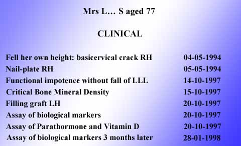
3 - Mrs L.S.
![]() A
small lady, aged 77 in 1994. She has suffered a great deal of misfortune
during her life, particularly during the war. After escaping the worst,
she has led a difficult life with great courage and a tremendous will
to live. Apart from numerous traumatisms, she suffers from cancer of the
blood which she bears with the same self-denial.
A
small lady, aged 77 in 1994. She has suffered a great deal of misfortune
during her life, particularly during the war. After escaping the worst,
she has led a difficult life with great courage and a tremendous will
to live. Apart from numerous traumatisms, she suffers from cancer of the
blood which she bears with the same self-denial.
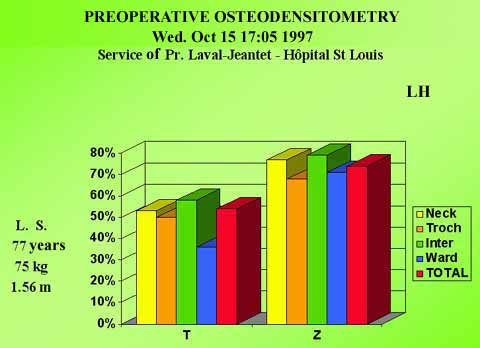
![]() In
1994, she fell and fractured the neck of her right femur. A one-piece
nail-plate was inserted. In mid-October 1997 she suffered from pain in
her left hip and displayed total functional impotence. A densitometry
was performed five days later. This revealed major osteoporosis with a
bone mineral loss of almost 50%, and more than 60% in the weakest part.
In
1994, she fell and fractured the neck of her right femur. A one-piece
nail-plate was inserted. In mid-October 1997 she suffered from pain in
her left hip and displayed total functional impotence. A densitometry
was performed five days later. This revealed major osteoporosis with a
bone mineral loss of almost 50%, and more than 60% in the weakest part.
![]() A
filling graft of natural coral was performed on the 20th of the same month.
Biological assays were performed during her hospitalisation and these
showed primary osteoporosis, in other words there was no disease or hormonal
cause likely to alter bone calcium.
A
filling graft of natural coral was performed on the 20th of the same month.
Biological assays were performed during her hospitalisation and these
showed primary osteoporosis, in other words there was no disease or hormonal
cause likely to alter bone calcium.
![]()
For further information: biological markers...
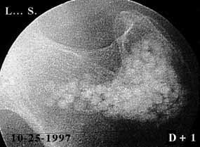
![]() X-rays
were taken just over a year later. It was noted that the coral had disappeared.
X-rays
were taken just over a year later. It was noted that the coral had disappeared.
 |
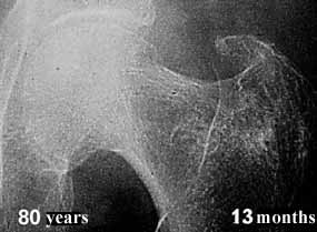 |
| Postoperative D+1 Coral graft visible |
Postoperative 13 months Graft totally disappeared |
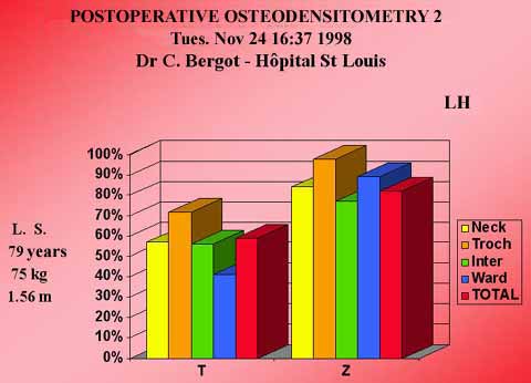
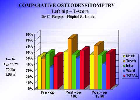
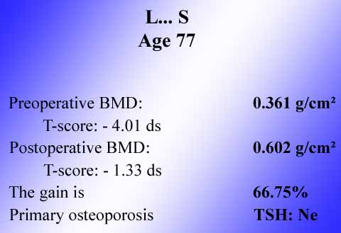
![]() A
new densitometry showed an increase of more than 65% in BMD in the grafted
sector.
A
new densitometry showed an increase of more than 65% in BMD in the grafted
sector.
![]()
![]()
![]()
| Previous page | Next page |