Revelation of bone loss
What does the lens see in an osteoporotic bone?
| Video section: JOURNEY TO THE CENTRE OF AN OSTEOPOROTIC BONE |  |
![]() First
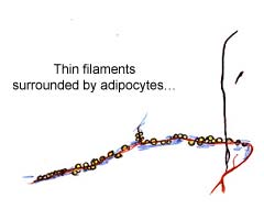
of all it sees nothing or rather it sees red. Mobile particles must be
washed out without mechanical force, these resulting from and being the
consequence of bone destruction. They are essentially fat cells, as has
already been said. It bears repeating because it really is fat. It is
easier to understand how falling even a short distance can lead to considerable
bone lesions…
First
of all it sees nothing or rather it sees red. Mobile particles must be
washed out without mechanical force, these resulting from and being the
consequence of bone destruction. They are essentially fat cells, as has
already been said. It bears repeating because it really is fat. It is
easier to understand how falling even a short distance can lead to considerable
bone lesions…
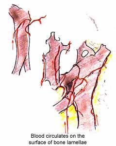
The field begins to clear and then, under the effect of low pressure lavage,
more solid structures – less fragile because of their connections
– start to appear.
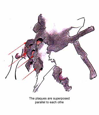
First of all we see absolutely white structures. These are bone lamellae.
They are single or connected to each other, forming plaques which are
positioned on top of each other.
 |
 |
![]()
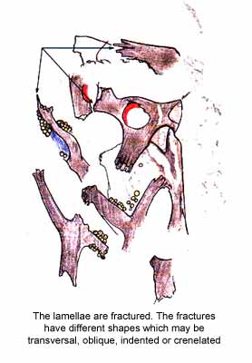
The lamellae or plaques are often fracture sites
 |
Different mechanisms may cause different types of fracture | |
| Chemical | - Variation in pH - Variation in 02 and/or CO2 levels - Release of proteolytic enzymes |
|
| Mechanical | ||
| Biological | - Macrophage action | |
On several plaques, an oval shape
can be seen, reducing |
|
 |
On each of the two mineralised plaques, perforated
with an even circular hole, two lacunae with uneven edges can be
seen, thinning the plaques which are almost transparent. |
![]()
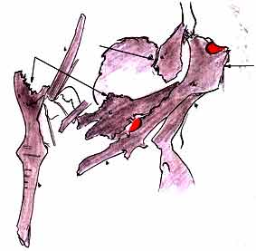
| On progressing, non-functional vessels
may be seen, |
|
 |
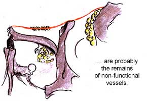 |
These are functional vessels, attached
to bone lamellae, |
|
 |
 |
![]()
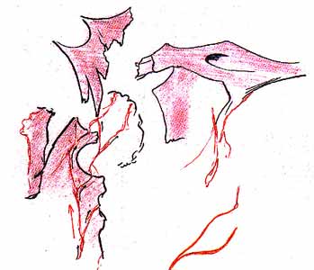
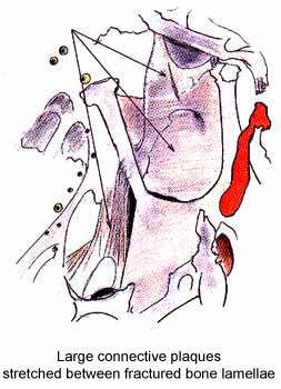
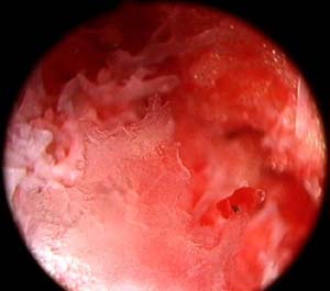
In other places, you can see soft tissue, either in the form of fine strips stretched between two lamellae
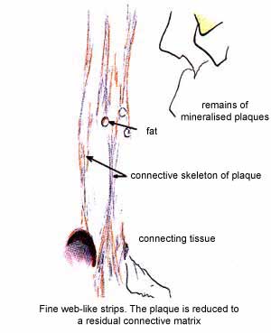
or in broad strips or plaques, perforated or not.
 |
 |
![]()
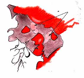
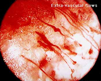
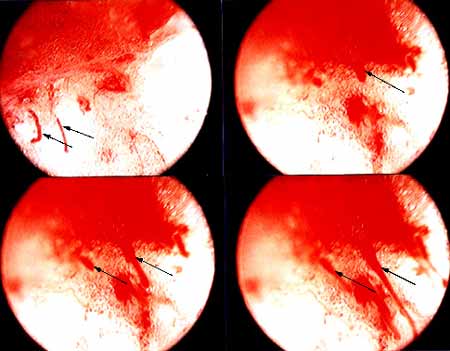
| You can see fluid of different viscosity from blood flowing from these broken vessels. |
 |
 |
![]()
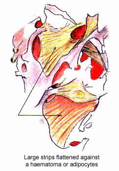
| These flows will create haematomas, |
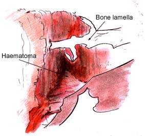 |
| The disappearance of bone lamellae may be due to:
|
 The micro-thrombosis can be seen as the black spot on the bone lamella |
![]()
| Previous page |